
Steady State Visual Evoked Potentials uses different frequencies of visual stimulii to distinguish between objects in BCI applications and have relatively high information transfer rates.
Evoked potentials are most commonly used EEG signals in BCIs. To recap, an evoked potential is a stereotypical response elicited when a subject is presented with stimulus. In the previous blogposts, we looked a P300 evoked potential, which is elicited when the subject encounters a rare stimulus within a stream of frequent or routine stimuli. In addition to this P300 potential, other types of potentials such as 1) visually evoked potential (VEP) generated by flashing lights, 2) steady state VEP (SSVEP) generated by repeated visual stimulus, 3) auditory evoked potentials (AEP) generated by clicks or tones (i.e., auditory stimuli) and 4) somatosensory evoked potential (SSEP) caused by somatosensory stimulation can be used in BCIs. In this blogpost we will focus on SSVEPs.
SSVEP-based BCI
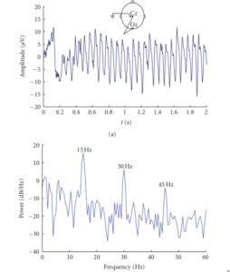
The idea behind using SSVEP in BCI applications is rather simple. Consider a scenario where the user has to order a beverage and there are three choices. Each of the choices can be represented as a visual stimuli, for example using smileys on screen flickering at different frequencies. The user then just has to focus on his/her choice (e.g., by looking at it). Focusing the attention on the button (which represents the user’s choice of beverage) flickering at a particular frequency will generate EEG signals (in the occipital regions) oscillating at the same frequency! This EEG signal, which oscillates at the same frequency as the flickering visual stimuli (and its harmonics) is known as the steady state visually evoked potential (SSVEP). The figure below shows a schematic of a typical SSVEP-based BCI.
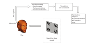 Figure 1 : SSVEP-based BCI [1]
Parameters affecting the performance of SSVEP-based BCI
The choice of several parameters can affect the performance of SSVEP-based BCIs [1]. First, the type of stimuli has been shown to affect the strength of the SSVEP response. Three main types of stimuli are used – 1) Light stimuli (e.g. LEDs, fluorescent lights), 2) Single graphics stimuli (e.g. rectangle, arrows) and 3) Pattern reversal (e.g. checkerboards). Both light and pattern reversal stimuli have been shown to produce a stronger SSVEP response than single graphic stimuli modulated at the same frequency [2,3]. Also, light stimuli produces a larger SSVEP response compared to pattern reversal, also resulting in the highest information transfer rates for SSVEP-BCI compared to the other two types of stimuli [4].
Most studies comparing types of stimuli have held constant most of the other variables, including color, frequency of the flickering stimuli and/or luminance of the stimuli, which could also affect the performance of the BCI [1]. However, from a practicality point of view it is easier to use a computer screen (i.e. reversal pattern or single graphics) than build dedicated hardware units to generate light stimuli. The downside of this is that the range of frequencies that can be used for visual stimuli is limited by the refresh rate of the computer screens.
The stimulus frequency is another parameter, that can be classified into low (1-12 Hz), medium (12-30 Hz) and high (30-60 Hz) frequency bands. Most of the applications have used low and medium frequency bands, however they have some substantial disadvantages, as visual fatigue has been observed in the range 5-25 Hz. Also, one study has shown the occurrence of epileptic seizure in the 15-25 Hz range with pattern and reversal stimuli [5]. Another issue with low and medium frequency bands is their overlap with the alpha band (8-13 Hz), which could result is high false positives. However, low and medium frequency bands produce higher amplitude EEG signals [4] compared to the higher frequency bands, which makes them attractive. Several approaches have been proposed to mitigate the issue of weak SSVEP amplitudes at higher frequencies. First, there is less spontaneous brain activity at higher frequencies compared to the low and medium ranges. Second, the SSVEP response can be increased with spatial filtering (e.g beamformers), [6].
The color of the stimuli is another important parameter, although a systematic study on how exactly the color of stimuli affects the BCI performance is still lacking. It has been reported that red light, when modulated at 11 Hz, generates the strongest response, whereas for blue and yellow light, the response strength was relatively less dependent on the frequency choice and the strength was weaker compared to red light [7]. Presently, red, gray, black and white light are most commonly used.
In the next blogpost we will look at some of the common applications and limitations of SSVEP-based BCI.
References
- Zhu, D., Bieger, J., Garcia Molina, G., & Aarts, R. M. (2010). A survey of stimulation methods used in SSVEP-based BCIs. Computational intelligence and neuroscience, 2010.
- E. C. Lalor, S. P. Kelly, C. Finucane, et al., “Steady-state VEP-based brain-computer interface control in an immersive 3D gaming environment,” Eurasip Journal on Applied Signal Processing, vol. 2005, no. 19, pp. 3156–3164, 2005.
- Z. Wu, Y. Lai, Y. Xia, D. Wu, and D. Yao, “Stimulator selection in SSVEP-based BCI,” Medical Engineering and Physics, vol. 30, no. 8, pp. 1079–1088, 2008.
- D. Regan, Human Brain Electrophysiology: Evoked Potentials and Evoked Magnetic Fields in Science and Medicine, Elsevier, Amsterdam, The Netherlands, 1989.
- R. S. Fisher, G. Harding, G. Erba, G. L. Barkley, and A. Wilkins, “Photic- and pattern-induced seizures: a review for the epilepsy foundation of america working group,” Epilepsia, vol. 46, no. 9, pp. 1426–1441, 2005.
- O. Friman, I. Volosyak, and A. Gräser, “Multiple channel detection of steady-state visual evoked potentials for brain- computer interfaces,” IEEE Transactions on Biomedical Engineering, vol. 54, no. 4, pp. 742–750, 2007.
- Y. Wang, X. Gao, B. Hong, C. Jia, and S. Gao, “Brain-computer interfaces based on visual evoked potentials: feasibility of practical system designs,” IEEE Engineering in Medicine and Biology Magazine, vol. 27, no. 5, pp. 64–71, 2008.