The EEG measures aggregate electrical activity across millions of cells but there are many complexities to consider.
The EEG had a tumultuous beginning – no one believed it was real (read Decoding the Electric Brain for its curious history). Yes, it’s an electrical signal from the brain, but how is it generated and what of the brain’s inner working does it allow us to see? To get to this we need to start with the basics.
The brain is made up of two primary cell types called neurons and glia that together form the jelly like folded tissue of our brain (grey matter, white matter, all of it). All this brain tissue is sitting in a very small volume of cerebrospinal fluid (CSF), which is basically salt water (sodium chloride or NaCl), but has other ionic salts (potassium, calcium, magnesium) as well as glucose and other nutrients. What this means is that CSF is a good conductor of electricity.
Within the tissue, Neurons are the active conductors of electrical signal, extending out long cables called axons that form connections with other neurons, along which they can propagate an electric signal in a non-attenuating manner. They do so by a process of exchanging rapid spikes of charge with the external fluid (the CSF) down its length. When the signal or spike has reached the end of the cable where the neuron connects with another neuron (the synapse), it triggers a release of chemicals, which in turn activates an inflow of electric charge in the receiving cell.
The cables that receive signals are called dendrites and are constructed differently from axons. They don’t have the insulating material needed for fast spike propagation. These currents on the receiving end – called synaptic and dendritic currents – are much slower and longer. Electrodes placed on the cell surface or within the tissue (done frequently in tissue in a dish) can easily measure both the spikes and synaptic currents.
However, while the electrical activity of neurons is probably the most studied aspect of brain activity, it may only be part of the story. Glial cells can sense the levels of activity in the milieu and modulate it by changing how much or how neurons can communicate with one another. More recently, it is becoming apparent that they also fire their own kinds of spikes that have nothing to do with neuronal activity1. Long sidelined, these cell types outnumber neurons in the brain 4:1 and may have far more of a story to tell than we know.
To complicate it even further, it has recently been shown that human cerebrospinal fluid contains various neuromodulating molecules (some yet unknown) that can alter the overall levels of excitability of brain cells2, so the influence goes both ways.
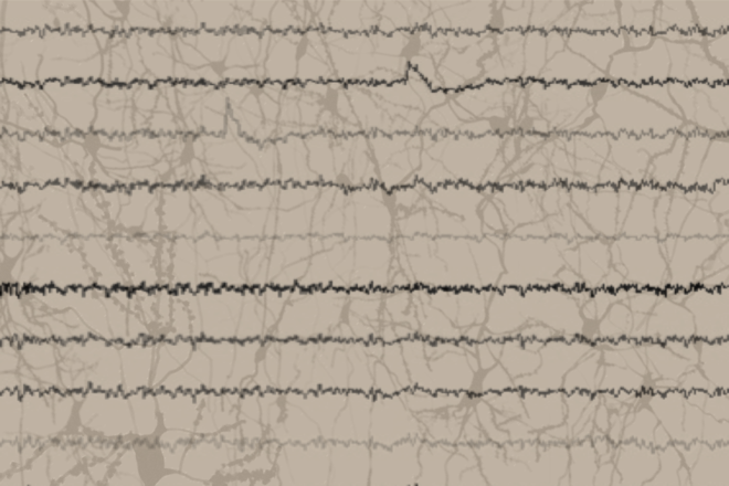
In the EEG we are not measuring activity of individual cells but the aggregate spikes and synaptic currents generated by whole swathes of tissue that contain millions of cells. What we measure are entire concerts of activity, with signal amplitudes that roughly reflect how many cells are actively communicating at any given moment or acting in concert with one another. This could be thought of as analogous to tracking the aggregate flow of activity on the internet, where you can’t read any individual email or piece of data but could understand some macro traffic patterns. That said, the measured signal also depends on the geometry of the tissue and the thickness of white matter and grey matter so it cannot be considered simply a direct reflection of aggregate neural activity.
So what then is the EEG? We are still learning. Even as we may look at the individual elements of the brain to try and understand how it gives rise to this aggregate signal, there are patterns and structures in this aggregate field signal that are difficult to explain in this bottom up way. Rather, we might think of it as a more holistic view of brain activity, a composite energy signature that could in turn give us insights into the underlying interplay that must take place within.
References
1Rungta RL., Glia Aug 2016
2Bjorefeldt A et al. J. Physiol Jan 2015