The human brain, encased within the skull, is the most protected and shrouded of our organs. Here are seven ways to probe and peer into it.
 Computerized Tomography (CT) is an upgraded version of the old X-ray technique where very powerful x-ray emitters rotate around you producing pictures of various angles and depths. With sophisticated computing these images are constructed into a three-dimensional image. Given that the white and grey matter of the brain possess different densities, it is possible to distinguish between them using a CT-scan. In the scan, white matter appears darker than grey matter because it is relatively less dense. It is useful as pathological processes such as tumors, lesions and tangles affect the density of grey and white matter. Of course, using X-rays comes with some disadvantages, such as the increased risk for developing cancer.
Positron Emission Tomography (PET) is a functional imaging technique used in medicine to observe metabolic processes for various purposes, detecting tumors being the big one. It involves injecting the subject or patient with a short-lived radioactive substance attached to a biologically active molecule (usually an analogue of glucose). The uptake of the active molecule by the brain tissue can therefore be observed by measuring the positron emission from radioactive decay, providing a measure of differential metabolic activity. It is typically used simultaneously with a CT-scan, to obtain a three-dimensional image combined with the tracer imaging. It is particularly useful for tumor detection since tumors metabolize glucose very differently.
Single-Photon Emission Computed Tomography (SPECT) is very similar to PET and also requires the injection of a radioactive tracer but uses a gamma-ray emitting rather than positron emitting radiotracer. PET and SPECT and even CT are not without risks and therefore not something to do for fun despite the coolness of the images.
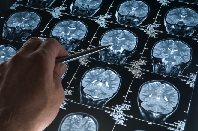
 Magnetic resonance imaging (MRI) is also a way to take pictures inside the brain but safer since it does not involve any radioactivity. MRI is based on nuclear magnetic resonance (NMR). This technique works by applying a pulse of radio waves under the influence of an external magnetic field and observing the emission spectra of the atomic nuclei of (mostly) hydrogen atoms. Since different types of tissue will produce different spectra this measure is used to form an image of the body. It takes more time, and the process, which requires lying still within the closed encasing of the machine, can be claustrophobic.
Functional magnetic resonance imaging (fMRI) This is perhaps the most widely used technique in human neuroscience and extends the principles of MRI to go from a static view to one of the brain in action. It does so by measuring changes in release of oxygen in the blood (Blood-oxygen-level dependent contrast imaging, or BOLD), which produces differential magnetic resonance. The theory is that because neurons lack significant reserves of oxygen (which unlocks energy within the cell), when they fire and use up the reserves it triggers the release of oxygen from the blood. The BOLD signal therefore serves as a proxy for neuronal firing activity in the brain. A relative increase in the blood oxygen in a region is what is meant by the term ‘the brain lights up’.
 Magnetoencephalography (MEG) measures the small magnetic field generated by the flow of charge in the neuron. While the magnetic field of individual neurons is extremely small, the collective fields of tens of thousands of neurons are detectable using SQUIDs or superconducting quantum interference devices. MEG is completely safe, can measure changes in neuronal activity on millisecond timescales (compare to ~1 second for FMRI) and also allow some resolution of activity below the surface of the brain (a drawback of EEG) making it perhaps the most powerful technique for imaging real time brain function. However, the catch is that to have low enough impedance so that they can pick up on these small changes in magnetic field these SQUID sensors have to be cooled to -276 degrees making it crazy expensive to use (~$3 Million for a single device and several hundred thousand dollars in annual running costs).
 Electroencephalography (EEG) measures the aggregate electrical activity in multiple regions of the brain using a set of scalp electrodes (typically silver chloride) with conducting gel to enable better contact with the skin (see Decoding the Electric Brain for its interesting history). Like MEG it can measure changes on millisecond timescales but does not have as good a spatial resolution and can only give you a surface view. This is, in part, because magnetic fields are not distorted by the skull and skin tissue whereas the electrical signal is. Nonetheless, it has an enormous advantage of being the least expensive of all of these technologies and still yields information that is of tremendous value and not yet fully explored. Its significant cost effectiveness (device costs ranging from $1,500 to $100,000), ease of use and portability (see taking Neurotechnology out of the Lab) make it possible to do many things with the EEG that simply cannot be done with any other technique.
And here’s a bonus. Its an 8th way really and the one you would never ever want unless the situation in desperate:
Electrocorticograph (ECOG) This is similar to an EEG but done directly on the surface of the brain to gain a much more fine scale signal resolution. Yup, your skull has to be cracked open and the dura opened up to place the electrodes. This is only done if you are undergoing neurosurgery and is used as a way for the neurosurgeon to map out functional areas (what happens when I stimulate this?) and more finely localize seizure activity so that they can be precise in their surgery.
Here is a quick view comparison
| Mechanism | Machine | Temporal Resolution | Spatial Resolution | Primary Use | Cost of Machine | |
|---|---|---|---|---|---|---|
| CT | X-rays taken at multiple angles | Lie flat and stil in a donut shaped openscanner | Single Time Slice (i.e. Static Image) | 1-4 mm | Structural Abnormalities | $70k -$1.5 Million |
| PET | Emission from injected radioactive tracer attached to a metabolic molecule (glucose) | Lie flat and stil in a closed scanner | minutes | ~1-4 mm | Detection of Tumor and other brain pathologies | $1.5 - $7 Million (depending on whether it comes along with CT or MRI) |
| SPECT | Gamma ray emission from injected radioisotop tracer | Lie flat and stil in a closed scanner | minutes | ~1 cm | Detection of Tumor and other brain pathologies | $400k-$600k |
| MRI | Nuclear resonance in response to radio wave pulses applied in a magnetic field | Lie flat and stil in a closed scanner | Single Time Slice | 0.2 to 0.5 mm2 x 1-2 mm | Structural Abnormalities | $150k - $3 Million |
| FMRI | MRI measurement of blood oxygen levels. | Lie flat and stil in a donut shaped scanner | 1-5 seconds | 1-5 mm voxels | Changes in blood oxygen following neural activity under different conditions | $500k to $3 Million |
| MEG | Magnetic signals measured using multiple SQUID sensors placed around the head | Sit still in chair with MEG machine surrounding your head | ~1 millisecond | 1-2 mm (including deep brain structures) | Changing neural activity profiles under different conditions | ~$2-3 Million; $200k annual running cost |
| EEG | Electrical signals measured using multiple electrodes placed on scalp | Scalp Electrodes, portable/ambulatory systems | ~1 millisecond | ~1cm (surface signal) | Changing neural activity profiles under different conditions, BCI, detection of Seizure and Sleep abnormalities | $1.5k to $100k |