There are many differences between mouse and human brains. How does this play out in the field potential signals such as LFP and EEG?
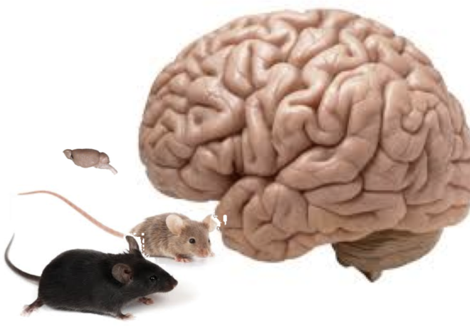
The vast majority of neuroscience research studies are conducted on rodents where the molecular and cellular physiology of neurons can be easily manipulated and measured. The overall architecture and cellular morphology appears to be largely conserved from mice to humans, which, at first glance, makes mice brains just look like much smaller human brains. However, increasingly, research is showing that there are enormous differences at the cellular level with implications for the nature of aggregate electrical activity.
Different Cell Types and Behaviors
Some of the biggest differences are of course, around glial cells. I’ve talked about this before here From Mouse Brain to Human Brain but just as a refresher, glial cells in humans are much bigger, control more neurons and produce many fold faster calcium spikes. There are also of course the new human specific Rosehip neurons that folks from the Allen Institute have recently described, a kind of GABAergic inhibitory neuron with a bushy tangled mess of a dendritic tree that occupies layer I of the cerebral cortex. These cells make homotypic gap junctions on the dendritic shafts of layer 2/3 pyramidal neurons and inhibit backpropagating pyramidal action potentials. Further, a study in human tissue by Gidon et al published last month in Science describes a new class of calcium-mediated dendritic action potentials (dCaAPs) in layer 2/3 pyramidal cells with a waveform and properties distinct from other known types of signals (might inhibition from rosehip cells have a role?). So altogether these distinctions between mice and humans in both neurons and glia come with specific alterations to how the flow of electrical activity is controlled.
Distinct Gene Expression
Beyond the cell types there are also the many differences in gene expression. A recent large-scale study of single cell transcriptomics (Hodge et al, Nature) from both human and mouse brains did show substantial conserved homology of cell types but also a great deal of divergence. The same group of genes typically identified similar cell types in humans and mice but beyond the gross similarities there were immense fine scale differences. This is not unexpected since this principle holds true even at the level of gross morphology and function, where all mammalian species have a similar architecture with roughly similar sensory apparatus, organs and body parts while the differences are in the details. The interesting aspect of this, however, is that many of the divergent genes were receptors and ion channels associated with connectivity and signaling. The serotonin receptors, for example, were found to be the second-most-divergent gene family. All this again points to differences in how electrical activity flows in the brain.
How does this play out in the overall electrical activity?
How does this all play out in terms of the aggregate structure of electrical activity that the mouse brain produces relative to humans? This is a million dollar question and one that has been inadequately explored. Between the different signaling in neurons and the glial calcium waves, and glial control of neurons, there must be something.
Having studied local field potentials in rodents and monkeys, one thing is clear is that there are many conserved features of the electrical signal. First, like the ECOG and EEG signals, LFPs measured in mice and monkeys also have a power spectrum with a 1/f – like decay pattern across the typically measured range of up to 50 Hz. Furthermore, the phenomenology of neuronal avalanches, a scale free structure of activity in the cortex, exists across species. That said, there are differences:
- While it is difficult to directly compare across field potentials recorded at different scales, the power spectrum appears to decay more steeply in rodents compared to monkey, and monkeys compared to humans.
- The alpha oscillation at ~10 Hz is most prominent in human recordings. You can see it a little bit in monkey LFP activity but not so much in rodents.
- The scaling relationship that characterizes neuronal avalanche dynamics (essentially a metric that describes the relationship between large size and small size propagating events), declines less steeply with lower temporal resolution in monkeys than rodents. Presumably this would be even more so in humans suggesting more robust spatio-temporal correlations in humans (see Petermann et al, PNAS).
- The waveforms in human EEG and ECOG are far more complex than in rodents – this is related to a steeper decay of the power spectrum.
Yet beyond these macro characteristics what else is different? This is still unknown and may hold the clues to some of the unique capabilities of humans.