Large intraperson variability in fMRI results for the same task questions the interpretation of decades of research, and makes a stronger case for EEG research.
EEG has long been the underdog technique relative to fMRI over the past few decades as money has poured preferentially into fMRI research and journals have been far more interested in publishing fMRI work than EEG.
see related posts EEG and FMRI Papers by the Numbers and EEG and FMRI Journal Stats
The simplicity of ‘this part lights up when you do X’ has made for easy PR around findings with very rudimentary conclusions like ‘if the same part lights up when you do Y as when you do X, then X and Y are functionally related’. This harkens back to two key assumptions:
1) the whole localization of function argument where function was thought to be localized to particular regions of the brain and
2) the argument that blood flow/oxygenation serves as proxy for activity and in turn ‘activity’ (by virtue of 1) dictates what exactly is being done in any region of the brain.
People are now increasingly opening their eyes to the challenges of this approach.
Localization of Function
The question of whether function is localized is a long standing one and has been debated since Broadmann started carving up the cortex into functional units in the late 1800s. Yet, while there is some localization simply by virtue of which part of the brain different sensory input arrives in, there has been a great deal of work over the last century demonstrating that memory itself is distributed and that the location of function in individuals are not identical and can even move around in an individual over time. The most compelling demonstrations come from the early work of Wilder Penfield which you can read about in this post: Stimulation, Sensation and Localization in the Cortex
The field of fMRI has for the most part ignored all of this.
Interperson Variability
First, there has been a fair bit of evidence in fMRI already that there is a lot of inter-person variability in which part ‘lights up’. Meaning people are doing the same task but different parts light up for different people, which already calls into question the whole assumption of some kind of absolute localization of function across individuals and therefore any interpretations of function based on location. The ‘average’ is not really meaningful if in reality it is just the number in the middle of substantial spread.
See related post The Myth of the Average Brain
In fact, people with half a brain – literally – can perform the same tasks that ‘on average’ light up on the side they don’t have.
See related post The Curious Outcomes of Neurosurgery
Intraperson Variability
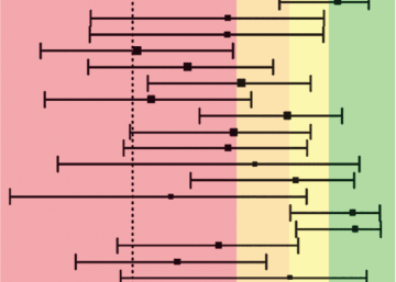
Then there is the aspect of intra-person variability. In a recent paper in Psychological Science titled What Is the Test-Retest Reliability of Common Task-Functional MRI Measures? New Empirical Evidence and a Meta-Analysis fMRI researcher Ahmad Hariri and his team at Duke University performed a meta-analysis to look at the correlations within individuals performing the same tasks at different time intervals – i.e. the test-retest reliability. The numbers were bleak. Across 64 different test-retest estimates (from papers where all results were reported), the average correlation was 0.397. Which meant that for the most part a different part was ‘lighting up’ each time. Hariri, who has been engaged in fMRI research for 15 years had this to say about his work in a recent article in Science Magazine. “This is my fault. I’m going to throw myself under the bus. This whole sub-branch of fMRI could go extinct if we can’t address this critical limitation.”
Nonetheless there were some interesting aspects to this:
First, the tasks that had the least reliability in general were tasks relating to emotion (most <0.2 correlation some as low as 0.02) and tasks relating to memory such as N-back memory tasks. On the other hand, the tasks with the highest correlations were a pain task (0.87, N=6), a sensory task (0.85, N=17) and motor task (0.84, N=17). Altogether this could be a reflection of a very distributed nature to emotion and memory (maybe this is the real story and fMRI research should focus on how the signal moves around), or a reflection of the tenuous relationship between the BOLD signal (blood oxygen) and function or both.
This brings us to this second point:
How does blood flow relate to function?
Making judgements about function based on blood flow might be a bit analogous to making judgements about what the world is up to based on where there are surges in power use. Yes, it has something to do with it, but a great deal happens at the same power levels and surges in power use could be completely unrelated to a particular task being measured. The location of BOLD hotspots moving may not even be evidence that function is even moving. If in fact there is no real ‘localization’ then more activity in a location is not necessarily an indication of ‘function’ since this logic presumes localization.
The EEG Advantage
This is where EEG has a potential advantage. It is not that there is not inter and intra person variability in the EEG. At least measured in spontaneous activity there is plenty.
See related post Intraperson Variability in the EEG
However, if there is no clear localization of function, then the most promising aspect is in the dynamical structure of activity. With its high temporal resolution there are many ways to explore this. In fact, if you are interested in the challenges around localization in the context of EEG, here is a Frontiers topic titled Does Electrical Stimulation Map Brain Function? that aims to get at the challenge of whether there is in fact functional mapping at all.
Beware the blanket verdicts. fMRI is not limited to blobology. It is also used for functionally more meaningful questions, like estimating the tuning parameters of neural populations and how they map onto the cortical surface (https://pubmed.ncbi.nlm.nih.gov/17964252/), which can be done with remarkable reliability (https://pubmed.ncbi.nlm.nih.gov/27620984/) and would be impossible to do with EEG. In my mind, cognitive neuroscience would gain a lot from a perspective which thinks about biological questions first and methods second. An argument about which one is better – EEG or MRI? – is about as meaningful as a debate about whether a screwdriver is ‘better’ than a hammer. It depends on what you want to use it for!
Varying reliability of fMRI is an additional layer of information. Emotion and short term memory are likely to be more malleable to experiences as opposed to sensory tasks – esp given that test-retest were done 4 months apart
I appreciate the clarity of the writing in this post; as a Clinical Health Psychologist who has practiced neuro and bio feedback for the past 40 years, I have appreciated the benefits of QEEG for the remarkable ways in which the temporal resolution of EEG offers additional metrics. We now have a much greater appreciation of “rich hubs” of activity thanks to EEG that help explain the 80% or better efficacy of neurofeedback. Glad I ran into this Sapien Labs email; thanks.