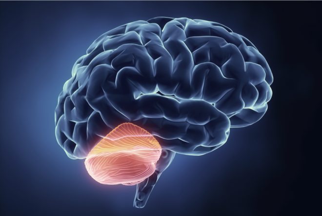
Long thought to be purely involved in motor activity, new roles have been discovered for the cerebellum in cognition. Yet so long on a backfoot in neuroscience research, the cerebellum is poorly understood.
The cerebellum has long been a misunderstood and often forgotten portion of the brain. But over the last decade or so, there has been increased interest in this unique and heterogeneous region which contains more than 4 times the number of neurons than the cortex itself, very few glia, and whose main output cells are inhibitory rather than excitatory. What’s more, as research has provided a greater understanding about how the cerebellum interfaces with different cortical regions of the brain, there has been a significant shift in thinking about what the cerebellum actually does.
Beyond motor control
Traditionally, the cerebellum was always seen as a region of the brain associated with the generation and control of movements. This arose from observations with patients that damage to the cerebellum caused noticeable disturbances in action and motor control. In addition, tracing studies of the time suggested that the cerebellum received inputs from multiple cortical regions (via the pons), but sent projections primarily to the motor cortex, via the thalamus. However, after the development of more sophisticated tracing techniques using neurotropic viruses, the multisynaptic pathways emerging from the cerebellum were more accurately mapped and led to the realization that the cerebellum sends projections to many different regions of the cortex (via the thalamus and dentate nucleus) including the prefrontal and parietal cortices.
 The Cerebrocerebellar Circuit: Input projections from the cerebral cortex first synapse on the ipsilateral pons and then cross to the contralateral cerebellar cortex. Output projections first synapse on the dentate then cross to synapse in the contralateral thalamus and finally project to the cerebral cortex. From Buckner, 2013.
In parallel, the number of cerebellar patient studies reporting cognitive disturbances led to researchers such as Jeremy Schmahmann to hypothesize in the early 1990s that ‘‘It may also transpire that in the same way as the cerebellum regulates the rate, force, rhythm, and accuracy of movements, so may it regulate the speed, capacity, consistency, and appropriateness of mental or cognitive processes’’. Since then, clinical studies have shown that cerebellar damage can impair cognitive processes such as task-switching, associative learning, and reasoning (see here for a review).
Similarly, with the advent in the late 1980s of brain imaging techniques such as positron emission tomography (PET), researchers started to reveal that the cerebellum became activated when participants were asked to perform cognitive tasks (e.g. involving language processing), rather than just motor ones (see here for a recent meta-analysis). Since then, it is common to see subregions of the cerebellum activated across a whole host of different cognitive tasks with techniques such as fMRI, but often these activations are only considered as incidental. To date there are still only a minority of imaging studies which have specifically targeted the cerebellum for a priori investigation, the most recent of which used resting-state functional connectivity MRI to map the pathways between cortex and cerebellum and suggested a strong functional role for the cerebellum in adaptive control of behavior and cognition.
see post 7 Ways to Peer into the Living Human Brain
Electrophysiology of the Cerebellum
In contrast to the increasing emphasis of the cerebellum within fMRI studies, the electrophysiology of the human cerebellum remains largely unexplored. One of the difficulties with measuring electrophysiological activity in the cerebellum is its poor signal to noise ratio compounded by the fact that there seems to be a greater level of individual variability in the cerebellum compared to the cortex (see Marek et al for details). This, along with the geometry and inhibitory nature of the purkinje cell (the main output cell of the cerebellum), as well as its location within the skull has typically led to the dismissal of EEG for measuring ongoing activity in the cerebellum, (although there are a couple of MEG studies which have reported cerebellar activity).
see related post Intraperson Variability in the EEG
Furthermore, there are only a handful of intracranial cerebellar studies performed in human patients (see here for a recent review) several of which have been performed several decades ago. Despite their rarity, they suggest that it is possible to measure task relevant modulations in spectral power, and that these modulations can be seen at higher frequencies (e.g. up to ~300Hz). Whether these frequency changes share any functional resemblance to the gamma band changes observed within the cortex remains to be seen. In addition, studies measuring event-related responses (ERPs) have suggested latencies of around 4 and 50 ms, and an absence of the longer-latencies more commonly observed in the cortex, again suggesting that oscillatory activity in the cerebellum may be distinct from that of the cortex.

At top, a spontaneous recording from the surface of the posterior vermis from a human patient, from Rétif (1964, Figure 4), reproduced with permission. Rétif speculates that the prominent wave between 70 and 90 ms had been evoked by sensory stimulation. At bottom, the spectrogram normalized with respect to the period before the large wave, 0–50 ms, expressed as percent change in power. Note the prominent sustained increase in power between 200 and 300 Hz, as well as more transient effects at lower frequencies.From Dalal et al 2013
Summary
In summary, the cerebellum has typically been on the backfoot of neuroscientific research, but is slowly emerging as a critical facilitator, or “corrector” of a wide range of cognitive and motor processes. In addition, it has been implicated in the emergence of several psychiatric disorders, including autism, schizophrenia, dyslexia, not only emphasizing its contribution from a clinical perspective, but also from a developmental one. Furthermore, working to develop new ways to tackle the complex challenges of recording electrophysiological signals, both invasively and noninvasively, from the cerebellum is a first step in potentially exploring, in more detail, the oscillatory activity of this curious region, and how this maps onto human cognition and behaviour.
References
Amaral, D., Schumann, C., & Nordahl, C. (2008). Neuroanatomy of autism. Trends In Neurosciences, 31(3), 137-145. doi: 10.1016/j.tins.2007.12.005
Andreasen, N., & Pierson, R. (2008). The Role of the Cerebellum in Schizophrenia. Biological Psychiatry, 64(2), 81-88. doi: 10.1016/j.biopsych.2008.01.003
Buckner, R. (2013). The Cerebellum and Cognitive Function: 25 Years of Insight from Anatomy and Neuroimaging. Neuron, 80(3), 807-815. doi: 10.1016/j.neuron.2013.10.044
Dalal, S., Osipova, D., Bertrand, O., & Jerbi, K. (2013). Oscillatory activity of the human cerebellum: The intracranial electrocerebellogram revisited. Neuroscience & Biobehavioral Reviews, 37(4), 585-593. doi: 10.1016/j.neubiorev.2013.02.006
Marek, S., Siegel, J., Gordon, E., Raut, R., Gratton, C., & Newbold, D. et al. (2018). Spatial and Temporal Organization of the Individual Human Cerebellum. Neuron. doi: 10.1016/j.neuron.2018.10.010
Nicolson, R., Fawcett, A., & Dean, P. (2001). Developmental dyslexia: the cerebellar deficit hypothesis. Trends In Neurosciences, 24(9), 508-511. doi: 10.1016/s0166-2236(00)01896-8
Petersen, S., Fox, P., Posner, M., Mintun, M., & Raichle, M. (1988). Positron emission tomographic studies of the cortical anatomy of single-word processing. Nature, 331(6157), 585-589. doi: 10.1038/331585a0
Schmahmann, J. (1991). An Emerging Concept. Archives Of Neurology, 48(11), 1178. doi: 10.1001/archneur.1991.00530230086029
Strick, P., Dum, R., & Fiez, J. (2009). Cerebellum and Nonmotor Function. Annual Review Of Neuroscience, 32(1), 413-434. doi: 10.1146/annurev.neuro.31.060407.125606
Timmann, D., & Daum, I. (2007). Cerebellar contributions to cognitive functions: A progress report after two decades of research. The Cerebellum, 6(3), 159-162. doi: 10.1080/14734220701496448