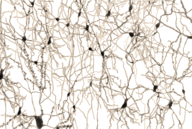
Much of neuroscientific research on brain signals is focused on studying the spiking activity of neurons in a dish. Can this really tell us how the brain works?
In studying electrical activity in the brain there are two distinct approaches. The first focuses purely on studying spiking activity of individual neurons while the second focuses on a complex aggregation of the activity of fields of cells including neurons and glial cells and the various electrical components from spiking activity to synaptic, dendritic and glial currents. The choice of one approach over the other has multiple considerations. The first approach of measuring neuronal spiking activity makes two deliberate assumptions: first that neuronal spikes are fundamentally more important than other aspects of electrical activity, and second, that the working of the brain can be inferred from these spikes. In contrast, focusing on field activity gives up the ability to distinguish the contributing elements [1,2] but nonetheless assumes that the aggregate electrical activity holds meaningful information. Arguments may be made for both approaches.
Neurons alone?
A great deal of the literature has been dedicated to identifying correlates between stimuli and behavior and neuronal spiking, particularly in rodents and monkeys. On the other hand there is also increasing evidence that glial cells play a significant and fundamental role in modulating and shaping the signal, particularly in humans where glia also give rise to much faster calcium waves and are much more abundant than in the rodent brain (glial: neuron ratio of 1.4x in humans compared to 0.4x in rodents) [3]. Therefore, neurons in isolation may contribute only one part of the story. Furthermore, there are practical considerations of measurement as well.
see related post Einstein, Astrocytes and EEG
From Elements to Aggregate
Consider the analogy of a simpler system of atoms and molecules that make up any particular type of conducting material. You could argue that the nature and state of the material could be best understood by measuring the changes in the energy and spin states of the electrons in each atom over time. However, this is extraordinarily difficult, if not near impossible, to accomplish from a technical perspective. Furthermore, it is not clear if a macro feature, such as conductivity, could actually be easily inferred by measurement of the electron states in individual atoms. On the other hand, measuring conductivity of the aggregate material with a simple conductivity meter does a pretty good job of estimating what is going on in the conductor and can ultimately be put more easily to use. Similarly, measuring the activity of individual neurons is more technically difficult. Besides requiring invasive implantation, microelectrode arrays today can typically only handle up to 100 electrodes while new generation technologies can measure a few thousand [4]. However, these are still only a tiny fraction of the brain’s billions of neurons. Therefore, analogous to the conductivity meter, field potentials might similarly be more easily exploited to gain insights into the brain’s behavior.
Mouse to human
Research in a reductionist framework that focuses specifically on neuronal activity is largely carried out in rodents and monkeys. Furthermore, the largest research spending is on experiments carried out in vitro. In vitro preparations are particularly amenable to measuring electrical activity of individual neurons. However, they all require sacrifice of the animal and therefore typically use rats and mice, or even invertebrates such as worms and fruitflies. This limits study to simple species. Consequently, our knowledge is largely about rodent signals and therefore cannot provide insight into the aspects of the signal that are unique to humans. Yet few papers in the literature qualify this, typically interpreting any results as universal to ‘the Brain’. For more on how the mouse brain differs from humans see related post From Mouse Brain to Human Brain.
Neurons to Brain
Notwithstanding all of the above, let us allow for a moment that neurons are singularly important and that mouse neurons are the same as human neurons. There are still perils in the dominant research approach that we use. Consider the following example of this much simpler system. Both diamond and graphite are constructed from the carbon atom. Diamonds and graphite cannot be formed from any atom other than the carbon atom and therefore the type of atom sets a constraint on possible outcomes. Yet diamond and graphite are completely different types of material. One is translucent, the other black. One is extraordinarily hard, the other soft. Indeed, important properties such as hardness and color are not properties of the atom itself but of the system overall. Therefore, one cannot ask if the carbon atom is black or hard. Rather, carbon alone can produce outcomes of completely opposite properties purely by virtue of how the atoms bond with one another. The nature of these bonds influences the way external energy such as light and mechanical forces interacts with it, and therefore the outcomes. Furthermore, the type of bonding is determined by the conditions of temperature and pressure of the environment in which they were formed. Consequently, studying the carbon atom in isolation or studying a carbon-based material formed under one set of conditions would not provide insight into the nature of an allotrope arising from a different set of conditions.
 Similar to how carbon atoms are predisposed to bond with one another, and do so based on environmental factors of temperature and pressure, the brain is characterized by the innate nature of neurons (and glia) to connect with one another in a manner that is based on their experience of the environment. It is therefore a reasonable assumption that the unnatural environment of the dish would alter some aspect of the way neurons wire up. Indeed, there is evidence that neurons have different gene expression patterns [5] as well as behave differently in vitro than in vivo [6]. A brain system where the formation of connections between neurons occurs under unnatural circumstances may therefore not provide insight into the working of the brain in its natural environment. The way external stimuli or electrical energy interacts with these preparations could also be fundamentally different. One would not want to unwittingly report the response of diamonds to light as those of graphite or vice versa. By this same logic studying the stimulus-response properties of neurons that are not wired in their normal environment may yield entirely misleading results.
See related post Brain in a Dish.
And with all this..
In vitro reductionist research can certainly give us some insight into general properties of the elements and their possibilities, such as the existence of plasticity, that communication occurs by synapses, and that signal can travel as action potentials. However, this is akin to understanding basic properties of atoms which don’t directly translate up to the understanding of the properties and behavior of different materials. Consequently it is grossly insufficient on its own. Indeed, considering all these aspects one has to wonder if the enormous body of in vitro scientific research is in fact, telling us anything at all about the functioning of the intact brain. And if it is, how this may translate to understanding of the human brain.
References
[1] O. Herreras, “Local Field Potentials: Myths and Misunderstandings,” Front Neural Circuits, vol. 10, p. 101, 2016.
[2] G. Buzsaki, C. A. Anastassiou, and C. Koch, “The origin of extracellular fields and currents–EEG, ECoG, LFP and spikes,” Nat Rev Neurosci, vol. 13, no. 6, pp. 407-20, May 18 2012.
[3] N. A. Oberheim Bush and M. Nedergaard, “Do Evolutionary Changes in Astrocytes Contribute to the Computational Power of the Hominid Brain?,” Neurochem Res, vol. 42, no. 9, pp. 2577-2587, Sep 2017.
[4] N. A. Steinmetz, C. Koch, K. D. Harris, and M. Carandini, “Challenges and opportunities for large-scale electrophysiology with Neuropixels probes,” Curr Opin Neurobiol, vol. 50, pp. 92-100, Jun 2018.
[5] P. R. LoVerso, C. M. Wachter, and F. Cui, “Cross-species Transcriptomic Comparison of In Vitro and In Vivo Mammalian Neural Cells,” Bioinform Biol Insights, vol. 9, pp. 153-64, 2015.
[6] S. A. Prescott, S. Ratte, Y. De Koninck, and T. J. Sejnowski, “Pyramidal neurons switch from integrators in vitro to resonators under in vivo-like conditions,” J Neurophysiol, vol. 100, no. 6, pp. 3030-42, Dec 2008.


I send congratulations to the authors of this article. Very good! I have been claiming the same for 20 years now. Two recent publications moving in the same direction:
https://www.researchgate.net/publication/315568664_The_Dynamical_Signature_of_Conscious_Processing_from_Modality-Specific_Percepts_to_Complex_Episodes
https://www.researchgate.net/publication/312938888_Astroglial_hydro-ionic_waves_guided_by_the_extracellular_matrix_An_exploratory_model
Good article.