This is a primer on fNIRS, what it does, the technical advances, challenges and potential applications.
With all the recent publicity around Kernel’s new fNIRS flow 50, we thought this would be a good time to provide a little bit of a primer on fNIRS.
What does it measure?
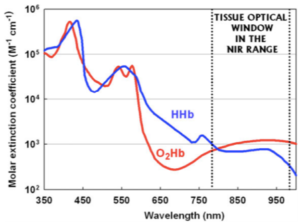
fNIRS which stands for functional near infrared spectroscopy uses infrared light to measure spatial changes in the oxygenation of blood. Generally, the more activity there is in any region of the brain (electrical and otherwise), the more oxygen it consumes so the idea is that by measuring the oxygen level changes you can measure how that map of activity changes over time. fNIRS works because the absorption of infrared light (~700 nm) is different between oxygenated and de-oxygenated blood – and its low energy enough that its totally safe.
 Different fNIRS devices exploit the signal in different ways. Time Domain or TD NIRS for example, pulses photons and measures the time difference in their arrival at the detector through the tissue, Continuous Wave or CW NIRS emits a continuous infrared signal and measures the changes in intensity. Essentially fNIRS can do a lot of what fMRI can do (which uses a whole different method of magnetic resonance to measure blood oxygen changes) but cheaper. Its also portable so can be used under more natural circumstances.
see related post 7 ways to peer into the living huamn brain
The technical advances
Technically, from an engineering perspective, the goal of companies like Kernel and others is to enhance the spatial and temporal resolution of the system (the smallest section of brain it can measure, how deep into the brain it can measure and how fast) while also increasing the ease of use and decreasing the cost. A lot of the advance is around how to position the source and detectors and how to pack them close together to cover more of the brain. Most fNIRS systems have spatial resolutions of ~ 1 cm, can go about 1.5 to 2 cm deep into the brain and have a temporal resolution of about 100 Hz. Kernel’s Flow has essentially miniaturized the whole thing without losing on any of the spatial or temporal resolution parameters.
The technical challenges
A big challenge in using fNIRS to map brain oxygen flow is getting good enough optical contact with the scalp without all the hair getting in the way and blocking the light. This is the same problem EEG has and tries to overcome with brush like electrode structures (in the case of dry electrodes). A second challenge is that people have different head shapes and optical path lengths differ on account of that making it difficult to be accurate for everyone. A third challenge (common to all neuroimaging methods) is dealing with artifacts caused by movement of the head. EEG can overcome this by using gel electrodes that stick in place, but it is tougher for fNIRS where it requires complex mathematical acrobatics that rely on assumptions that are a bit of guesswork.
The REAL challenge
The much bigger challenge is what the spatial pattern of oxygen consumption in the brain can really tell you and what you can do with it. Enthusiasm for fNIRS rests on the assumption that knowing where there is activity is informative about what is happening. The challenge with functional localization is that it both exists and doesn’t – obviously whatever the brain is doing happens has to reflect in activity somewhere. But this somewhere could be a bit different for each person (see this post on functional localization), and it moves within a person (see this post on the huge intra-person variability in fMRI and how it has challenged years of research). Second, just because activity (here oxygen consumption) is happening somewhere doesn’t mean you know what it’s about.
Imagine if you measured a huge increase in food and electricity consumption in in Atlanta – that would signal more activity and you would know something was going on, but just not what was going on. But even assume that you do know that time, that it means the super bowl is going on because you were also looking at what was happening. Could you subsequently predict when the super bowl is going on just by measuring a change in activity in Atlanta? Not a chance. Next year it may move somewhere else, and besides an increase in activity in Atlanta could just as well be because of a convention. You get the picture. Consequently, many research studies that try to relate fNIRS activity in some region of the brain to different kinds of brain function (cognitive, social etc.) have pretty weak results – small differences at p<0.05, which from the perspective of any reliability of prediction at an individual level is pretty much useless.
All is not lost
While the grand promise of predicting what you are up to based on your blood oxygen may be a tough one to realize, one obvious and useful application is in monitoring recovery from any kind of brain injury or damage to the cortex – stroke for instance arises from a blockage of blood flow and damaged tissue uses less oxygen so you can tell by the increase in oxygen consumption if the brain region is recovering. There may also be other neurological and mental applications that identify more subtle blood flow inequities in the brain as underlying causes. There are also possibilities that arise out of looking at the combination of regions or ‘networks’ of blood flow which could likely tell you more than changes in individual regions but it is yet unclear The jury is out.