Pieces of brain in a dish can give rise to synchronous electrical activity with similar characteristics to that measured in the intact brain. What aspect of this activity is then truly meaningful in the context of the living, functioning organism?
What’s unique to the living, functioning brain? The brain is sculpted in an experience dependent manner but not all of its function is motivated by its external environment.
Understanding the behavior of brain tissue in the absence of stimulus can help us understand its intrinsic nature. My own interest in Neuroscience was sparked by the eerie fact that you can take a piece of brain from a (once) living animal and keep it alive in a dish for several hours and measure its electrical activity. What does this bit of brain do on its own and what does that mean?
Acute Slice
This technique dates back to the 1960s or 1970s. Called the acute brain slice it is generally performed with adult rats and mice. In one common protocol it involves anesthetizing them and making the heart pump oxygenated artificial cerebrospinal fluid (CSF) into the brain to cool it down. The cooled down brain is then removed from the animal and cut into very thin slices of about 100 to 300 microns while it is submerged in the oxygenated artificial CSF. The slices have to be thin because the vasculature is no longer pumping blood so the cells have to survive by taking oxygen from the CSF they are submerged in – thicker than 300 microns and the oxygen in the surrounding fluid cannot penetrate the tissues and cells deeper in the tissue would asphyxiate. At this point the animal is dead but the brain is not, although it lives only in a bunch of pieces and not all as one integrated organ. The image below shows a rat brain in a dish, the blade of the vibratome positioned to slice it (with a bunch of already cut slices around it and finally, one of those slices in a dish ready for experimentation.

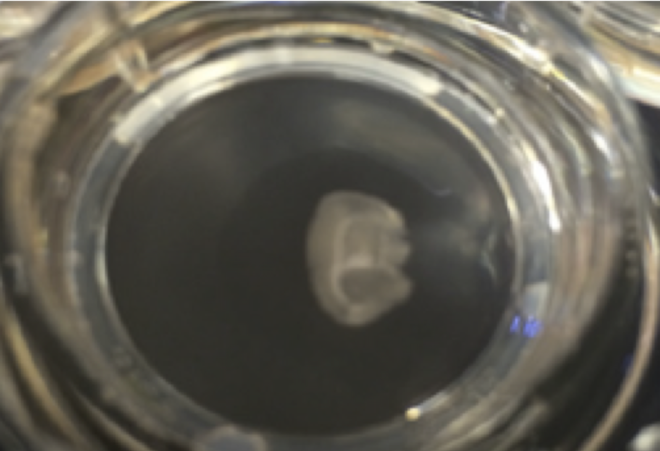
This is hard to do with larger mammalian species like monkeys not just because the animal is sacrificed in the process but also because the bigger the animal the harder the skull – which means that the process of extracting the brain would take too long for the cells to survive. Nonetheless with rats and mice, this is pretty easy and if you have stable hands and are swift in the extraction and delicate in handling of the tissue, the brain slices can live in a dish of oxygenated artificial CSF for ten hours and sometimes more. People have taken advantage of this to measure activity using multielectrode arrays, patch clamp electrodes and various imaging techniques. The image below shows an acute slice placed on a multi electrode array (from Beggs and Plenz, 2003, left) and a close up image of an acute brain slice through the microscope.

 Just like the living brain, acute slices produce activity even in the absence of any kind of stimulus – sensory input or otherwise. Neurons fire and there is synchronous activity measurable on arrays of electrodes with a statistical structure remarkably similar to the living brain (for examples see Beggs and Plenz 2003, Petermann et al, 2008, Thiagarajan and Plenz, 2010). These slices can carry both biochemical and electrical signatures of events experienced by the animal before the brain was extracted and sliced up. If this is so, are these slices recreating experiences and reminiscing in the dish?
see related post From Mouse Brain to Human Brain
Organotypic Slice Cultures
Of course, these are slices from adult animals that have lived several years and formed many memories. Another preparation that has a strangeness to it is the organotypic slice culture. This is perhaps even more remarkable than the acute slice. Here brain slices are prepared from new born mice (or rats) where the cells of the cortex have not migrated to their final positions yet. The slices can be placed in a dish where the cells will continue their migrations to their final positions, spreading out and forming a thin 2D layer of brain with an architecture very much like the living cortex. In this configuration they can live in an incubator in a nutrient rich medium for weeks. Unlike the acute slices these slices come to the dish without much ‘experience’ of the sensory world. The mice they come from are newborns that had not yet opened their eyes. In fact this technique can only work with newborn animals, so you wouldn’t expect much of this 2D slice of brain. Yet, even so, in labs such as that of Dietmar Plenz’s at the NIH where they have grown these slices on top of microelectrode arrays, there is robust activity. After about two weeks of just sitting quietly in the dark incubator without any intervention, not only are the neurons actively firing but the slice suddenly begins to develop a structured symphony of synchronized electrical activity – again with a structure similar to that in the living brain. If not a memory or representation of living sensory experience, what does this activity represent?
see related post Dendrite Complexity and Intelligence
Dissociated Culture
The intrinsic nature of brain cells to connect and generate electrical activity is most apparent in a preparation called dissociated cultures. Here unlike the organotypic cultures where whole slices of brain are grown in a dish, the cells are extracted from the newborn rat or mouse brain and put through an incredible assault to tear them apart from each other and sever their neurites. Of course, many cells die in this process but enough survive and these can be placed on a dish coated with a sticky nutrient rich gel. Common practice is then to inhibit the growth of glia so that they don’t overwhelm the neurons – a questionable practice given our growing understanding of their significance and influence on neurons (see Einstein, Astrocytes and EEG). Nonetheless the neurons in the dish will regrow their neurites which squirm around the dish seeking out other neurons to connect with. Over the course of a couple weeks the cells would have developed into a dense network. These are sparse networks of neurons and so the synchrony measured in the local fields of slices and cultures is not obvious, but the neurons are certainly actively firing and communicating. Again, this happens entirely in the absence of any kind of stimulus or significant prior sensory experience.
 Thus while various sorts of electrical and biochemical stimulation in the dish can cause plasticity and changes to the synaptic communication and electrical activity, the basic structure of activity can be created and maintained in its virtual absence.
This begs the question of what aspect of the electrical activity or signal is relevant in the context of the functioning organism. Are there fundamental differences between the activity in a dish versus the intact organism? Correlates may be many, but what is truly meaningful?