In EEG, voltages recorded at each electrode are computed with reference to another electrode. The choice of this electrode reference impacts a number of EEG measures.
Choice of the reference electrode location is one the most critical issues in EEG recording. It impacts not only the amplitude of the recorded potential but also its temporal structure. For example, consider the visual evoked potential (VEP) recorded from the occipital electrode O2 against electrodes placed in the mid-line (a), frontal (b) and temporal areas (c) [1].
 Figure 1: Taken from [1]
It can be seen that both the peak amplitude and the temporal structure of the evoked potential is dependent on the choice of reference, with the N100 (peak occurring 100 ms after stimulus) having the smallest amplitude (almost at the level of noise!) for the reference electrode in the temporal area.
An ideal reference location should be electrically neutral, which is not the same as being at the farthest from the EEG generators. A reference location should also take into account the size of the EEG generator region (which is approximately 10 cm^2). In the example above, although frontal area is farthest from generators of VEP which are in the occipital area, still the N100 amplitude is smaller compared to the mid-line reference. Thus it is not enough to place a reference electrode at a place where you think there are no generators of EEG, but one should also account for how it electrically impacts the reference location.
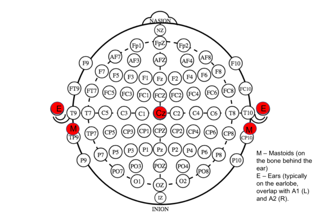
Typically used references include vertex (Cz electrode), nose, linked mastoids (behind the ear) or ears, common average reference and reference electrode standardization technique (REST) or infinity reference technique [2]. Since referencing is essentially a linear operation, irrespective of the reference electrode used during on-line EEG recording, the EEG data can always be re-referenced to any other electrode (either physical or virtual) for further processing (provided the hardware allows it – see practical considerations below).
Effect of reference choice on power spectra
Power spectral analysis is one of the most commonly used technique to analyze EEG data.
see related post Factors that Impact Power Spectral Density Estimation
Hence the impact of the choice of reference on this measure is important to be understood. Yao and colleagues compared four reference techniques – left mastoid, linked mastoids, average reference and infinity reference on the power maps computed in alpha band for eyes closed EEG data [3]. As it can be seen in the figure 2 below, the topography looks distinct for each reference. Statistically significant differences were observed between the total power (in Alpha-1- (7.5-9.5Hz) and Alpha-2 (10-12Hz) bands) over all channels for the four reference techniques, with the total power for average reference being the smallest and mastoid reference being the highest.
 Figure 2 : Taken from [3]
Effect of reference choice on connectivity measures
It has been shown that commonly used functional connectivity metrics (at sensor level) such as coherence, phase locking value, phase lag index are also confounded by reference issues. The fluctuations in the amplitude of the reference signal have significant impact on phase synchrony measures, while the fluctuations in the reference signal power affects coherence computation [4]. Also, usage of a common reference channel (like Cz for example) can lead to spurious functional connectivity estimates [5]. This is illustrated in figure 3, where fluctuations in reference potential leads to spurious coherence. Using separate reference R1 and R2 for each of the electrodes or bipolar derivation can be used to solve the common reference problem.
 Figure 3 : Taken from [5]
Impact of reference on machine learning outcomes
Apart from connectivity analysis or power spectral analysis that is performed on EEG data, the use of machine learning techniques to classify EEG signals is gaining popularity. This is extremely useful in clinical settings, particularly in epileptic EEG data, where the task is to classify the signals into pre-ictal, interictal and post-ictal segments. Given that different types of reference systems are used in clinical data, the performance of such algorithms can considerably vary which is undesirable in clinical application.
Lopez et. al compared linked mastoid and average reference using hidden Markov Model classification system on Temple University Hospital (TUH) EEG corpus [7]. Their results showed that a system trained on linked mastoids reference data outperformed average reference for this dataset. They also showed that compared to linked mastoids, the performance of the algorithm was lower when both linked mastoids and average reference was used. To reduce the bias between these two reference systems, they implemented Cepstral mean normalization, a technique commonly used for speech signals. However, this decreased the performance of the classification algorithms even further. Although it is critical that some form of channel normalization should be done in order to maintain performance across different reference choices, this does not seem to be a trivial task.
Practical Considerations
Any choice of reference will bias the analysis and should therefore be accounted for while interpreting the result. Today many hardware systems provide pre-referenced outputs for channels, rather than allowing a flexible selection of reference or re-referencing of the data. Consequently, this is a fundamental consideration in selecting the device you use. While some references may perform better for some outputs compared to others, there appears to be no ‘best reference’. It is important however to be consistent in the referencing in comparing results or aggregating data from different sources.
References
- 1. Nunez, Paul L., and Ramesh Srinivasan. Electric fields of the brain: the neurophysics of EEG. Oxford University Press, USA, 2006.
- Yao, D. (2001). A method to standardize a reference of scalp EEG recordings to a point at infinity. Physiol. Meas. 22, 693–711. doi:10.1088/0967-3334/22/4/305
- Yao, Dezhong, et al. “A comparative study of different references for EEG spectral mapping: the issue of the neutral reference and the use of the infinity reference.” Physiological measurement26.3 (2005): 173.
- Hu, Sanqing, et al. “On the recording reference contribution to EEG correlation, phase synchorony, and coherence.” IEEE Transactions on Systems, Man, and Cybernetics, Part B (Cybernetics)40.5 (2010): 1294-1304.
- Bastos, André M., and Jan-Mathijs Schoffelen. “A tutorial review of functional connectivity analysis methods and their interpretational pitfalls.” Frontiers in systems neuroscience9 (2016): 175.
- Chella, Federico, et al. “Impact of the reference choice on scalp EEG connectivity estimation.” Journal of neural engineering13.3 (2016): 036016.
- Lopez, Silvia, et al. “An analysis of two common reference points for EEGS.” 2016 IEEE Signal Processing in Medicine and Biology Symposium (SPMB). IEEE, 2016.
A number of diagram errors. A1 is left ear and A2 is right ear. (odd nuimbers left and even numbers right side).
Mastoid electrode formal placements are not well agreed upon but CP10 and TP9 are not R-L homologs. Error in part because you also show TP7 and CP8 as L-R homologs in error. Also FC7 and FT8 are also not homologs.
You do not describe the physical theory behind forming the common average reference and the errors introduced when limited electrode placements and/or numbers violate the theory behind the CAR.
Also you do not discuss the Laplacian reference which, if done properly, is the best “montage” as the Laplacian is the most localizing montage technique (see Paul Nunez book).
Finally you do not discuss the issue of electrode number (more is not always better). E.G. there is the issue of saline short circuiting (bridging) activity between closely spaced adjacent electrodes (compared to use of thicker paste) thereby messing up coherence measurements. AND LOTS MORE.
There would a real service to researchers to produce an electrode ‘standards’ document.
Frank
Thanks for the catch on the diagram and writing in! We will fix them shortly. There are a number of aspects to discuss of course and can’t easily be covered in a short blog post. We will try to cover some of these as best we can in a subsequent posts. Agree that an electrodes standards document would be great to have but is very difficult to implement given the range of configurations and options that in many cases are built into the hardware.
Thanks for the comment. Most of the concerns you raise has been addressed and will be published as next blogpost. However, it is debatable whether surface laplacian technique being the ‘best’ montage, as it can easily miss deep/distributed sources and works well only with shallow ones. More on this in the coming blogpost!