Astrocytes shape key features of the EEG signal and may be crucial for effective cognitive processes.
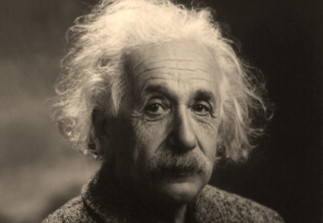
In 1955, Dr. Thomas Harvey acquired a brain. Whether he stole it or rightfully acquired it is a point of contention, but what he did with it is not. He sliced it up, preserved it in celloidin, posted 12 two-hundred-slide samples in the mail, and placed the rest into two jars. When Dr. Harvey moved, the brain moved. He stored it first in his basement, and later in a cardboard box under a beer cooler in his office. This is where it was when a young journalist came looking for Einstein’s brain in 1978.
1978 was a better year for Einstein’s brain than 1955. In response to the 12 samples Dr. Harvey had mailed to scientists in 1955, he had received few responses. Those who had studied the slides saw nothing particularly special about the brain. When word of Dr. Harvey’s preserved treasure hit the press in 1978, scientists were interested. Samples were sent and instead of radio silence or news that the brain was indeed unremarkable, scientists found differences.
An uncommonly high ratio of glial cells to neurons was one of the differences reported. Astrocytes and oligodendrocytes, collectively known at glial cells, have long been thought of as the brain’s housekeeping cells, simply cleaning up while neurons do the ‘real’ work. Though the study presenting Einstein’s unusual glial to neuron ratio is not without critics, today there is further evidence that astrocytes are much more than support cells.
Astrocytes and EEG
EEG records aggregate fields of activity in the brain, generally thought to be signals generated by neurons. Yet the phenomenology such as oscillations (see more about Alpha Oscillations here) that are picked up by the EEG in the field have been difficult to explain despite valiant efforts at simulating networks of neurons. (See related post What does the EEG Signal Measure?). Oscillations in the EEG, both in the alpha and gamma, frequency bands, have been associated with attention and there is a vast literature showing correlations between their occurrence and cognitive tasks requiring attention and memory.
What role do astrocytes play in generating gamma oscillations measured by EEG? Dr. Terrence Sejnowski and colleagues at the Salk Institute set about trying to find out (see the study here). Measuring activity in local fields in mouse brain slices in a dish along with imaging of calcium signals from astrocytes, they found that gamma oscillations in the local field were preceded by calcium signaling in the astrocytes. But does it have any relationship to the EEG, which is measured at a very different resolution? To answer this question they created a transgenic mouse that expressed a toxin that could selectively stop the astrocytes from signaling. This engineering came with a control feature that allowed them to enable and disable their ability to communicate with neighboring cells. With this the team set about measuring and comparing the EEG when astrocyte communication was turned on and off.
EEG in Mice
As you can imagine, recording EEG from a mouse is a bit more difficult than in humans given their size and the shape of their head. Consequently, this required surgical procedure. The mice had to be put under anesthesia, their heads shaved and the electrodes bolted on with screws and connected to a miniconnector soldered onto the mouse’s skull. After this rather difficult procedure however, they were able to measure EEG continuously while they controlled the behavior of the mouse astrocytes. The results were not spectacular but still, there was an effect. When astrocyte activity was turned off when the animal was awake, the power spectrum showed a reduction in the 20-40 Hz range (but curiously had no effect during sleep). In a surprising twist, the team also found that mice without communicating astrocytes were unable to recognize novel items in behavioral tests.
“This is the first time we have been able to do a causal experiment, where we selectively block gamma oscillations and show that it has a highly specific impact on how the brain interacts with the world,” Dr. Sejnowski explains. The experiment also shows that despite communicating at a slower speed than neurons, astrocytes are critical for some of our most important brain functions and play a role in shaping the EEG signal.
From Mouse to Einstein
The effect of astrocyte activity on the mouse EEG was modest at best. But Einstein was no mouse, and astrocytes may be even more crucial in shaping the features of human EEG that relate to cognitive processes than in mice (see related post From Mouse Brain to Human Brain). The diameter of a human astrocyte is 2.6-fold larger than in a mouse. They also have 10x more processes that connect to more neurons, and signal those same calcium waves that precede gamma oscillations in mouse 4X faster. Human astrocytes are also far more responsive to glutamate, a central neurotransmitter in the brain. On a molecular level, gene expression between human and mouse astrocytes differ much more than the gene expression between human and mouse neurons. It may be that the cognitive advances we have taken as a species are more dependent on our glial cells than neurons and the same experiment in humans (which you couldn’t really do) would deliver much more profound results in the EEG.
Perhaps Einstein’s glial to neuron ratio is worthy of notice after all.


Here are some related links regarding correlations of astrocyte EEG activity in the infra-low frequency range (below .1 hz). This technique was pioneered by Siegfried and Sue Othmer here in the U.S., but also related to Birbaumer’s slow cortical potentials work in Germany.
http://news.eeginfo.com/on-the-life-of-marian-diamond/
https://eeginfo.com/research/articles/David-Kaiser-ILF-Ultradian-Rhythms.pdf
Recent profile of Ben Barres at Stanford, Glial Guru.
http://www.the-scientist.com/?articles.view/articleNo/49226/title/Glia-Guru/