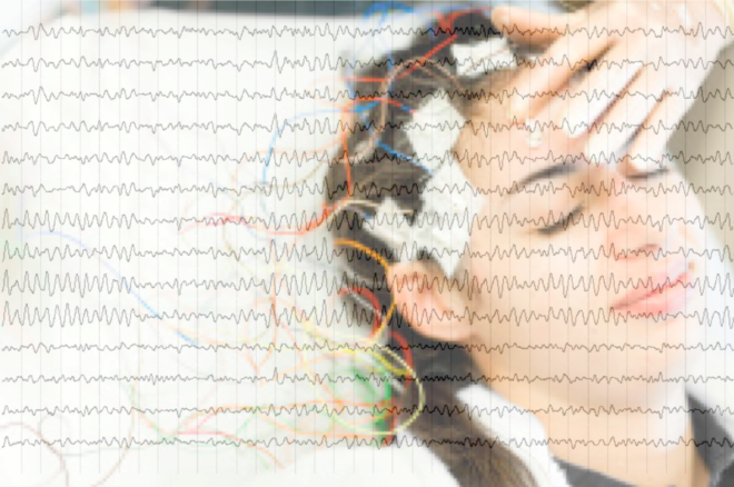
Activity in the alpha band has been a subject of much interest since the first recording of EEG. However methodological inconsistencies and confounding changes across a multitude of conditions obscure its meaning.
Ever since it was first shown on a brain recording trace in the early 1900s, EEG activity in the alpha band has been subject to considerable investigation by EEG researchers worldwide. It is often the dominant peak present on a frequency spectrum and demonstrates a stark increase in amplitude when a participant closes their eyes at rest.
The prevalence of alpha activity, perhaps unsurprisingly, has led to alpha increases and decreases being reported in a vast wealth of EEG studies employing both resting-state and task-based approaches, as well as in various clinical populations. But do all these studies join up? Are there patterns emerging that allow us to deconstruct the functional role of alpha in the brain, taking into account the broad spectrum of findings? Or is it just jumble of results? And importantly, as a crucial element to teasing out the relevance of alpha band activity, are the methodological standards utilized across these studies actually similar enough for us to meaningfully compare between them?
Resting state alpha changes
Focusing on one group of studies, namely resting-state EEG studies, allows us to determine the level of consistency, not only in methodological approach, but also in reported changes in alpha activity across different regions of the brain within a fairly constrained experimental set up (eyes open or eyes closed whilst at rest).
We ask here what kind of pattern emerges when comparing across the major brain disorders (e.g. ADHD, addiction, schizophrenia, depression, anxiety, OCD, autism, PTSD etc.) as well as mindfulness and meditation studies.
Differences in Methodological standards
The first thing that becomes obvious is that the methodological parameters used across studies and across research themes are not always consistent. While ‘alpha band’ activity may appear a simple standard the divergence in parameter choices shows that this is not so.
Methodological differences extend from selection of different referencing systems (see here for one discussion about different referencing approaches) to filtering of the signal using different frequency ranges, different epoch lengths and different algorithms, parameters and commands for estimating the power spectrum (see Factors that Impact Power Spectral Density Estimation for a discussion on what’s important to consider). An added challenge is the poor reporting in the methods sections about the specific parameters used for this spectral transformation, with some studies simply stating that they performed a fast fourier transform with few other details. The ability to draw meaningful comparisons between studies, which may potentially show different patterns of results, but which do not provide adequate method descriptions, is therefore limited.
Other sources of variability between studies include selecting different frequency windows to reflect the start and end of the alpha band window, reporting relative, not absolute activity, and log transforming data before statistical analysis.
| Variation in Methodological parameters across resting-state EEG studies. | |
| References | Mastoids, earlobes, nose, average brain, Cz |
| Low pass filter | Ranging from 22Hz – 300Hz |
| High pass filter | Ranging from 0Hz – 2Hz |
| Epoch length | Ranging from 1second – 60seconds |
| Spectral analysis (some examples) | pwelch, fft, psd or spectrogram.m; Hanning or Hamming windows with 0% to 75% overlap. |
| Alpha band start | Ranging from 6-8.3Hz |
| Alpha band end | Ranging from 11.5-14Hz |
| Sample size | Ranging from 14-407 |
Studies also vary according to the sample size, their selection criteria and meta-data reporting (see Human Brains and the Control Issue), and how well their clinical groups are “matched” (or not) to their healthy populations.
Although the precise parameters are often matched to the needs of the experimental dataset, the lack of standards in EEG research, and the mathematical complexity behind selecting the “best” parameters means that opportunities for improving consistency and reliability across studies are being missed.
Patterns? Or a jumble?
Early studies first labelled alpha activity as a sign of cortical idling but more recently it is thought to reflect the suppression of unwanted information or to facilitate communication between brain regions, especially when studied in relation to attentional processing (see Alpha Oscillations and Attention). What’s more, alpha changes have been reported across a wide range of clinical populations (and meditative states), potentially contributing their symptomatology.
But there is a relatively mixed patterns of results with this style of data analysis and reporting when you start to look across the spectrum of disorders which makes it difficult to build firm conclusions:
| Alpha increases across frontal regions
(vs a healthy control group) |
Alpha decreases across frontal regions (vs a healthy control group) |
| Patients with Schizophrenia (n=127, eyes open & closed) | Patients with Schizophrenia (n=26, eyes closed) |
| Adults with ADHD (n=100, eyes open) | Girls with ADHD (n=140, eyes closed, relative power)
Boys with ADHD (n=224, eyes closed, relative power) |
| Opiate users (n=42, combined analysis for eyes open and closed) | Opiate users (n=40, eyes closed)
Gambling addicts with high impulsivity (n=109, eyes closed) Daily smokers (n=52, eyes closed) |
| Adolescents with bipolar disorder (n=39, eyes open) | Individuals exposed to early life stress (n=407, eyes closed and open)
Patients with PTSD (n=170 eyes open) |
| During acem meditation vs rest (n=18, eyes closed). | During meditation vs rest (n= 71 eyes closed, relative power).
|
In summary, the range of studies reporting changes in alpha power across different regions of the scalp means it is difficult to make sense of it all. Employing pre-defined methodological standards and moving away from using arbitrarily segmented frequency bands in broad predefined regions, will help advance the field of EEG and what it has to offer from both a clinical and research perspective. However, more significant is that the confounding nature of these results calls into question the relevance of a single band based approach of reporting.
Ver useful and insightful article